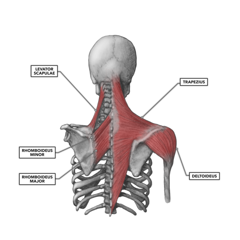
Shoulder Upper Back Anatomy : Shoulder Upperback The Yoga Lab - The extrinsic back muscles are located in the back, but act to produce movements of the shoulder and assist respiration.
Shoulder Upper Back Anatomy : Shoulder Upperback The Yoga Lab - The extrinsic back muscles are located in the back, but act to produce movements of the shoulder and assist respiration.. Upper right back pain under shoulder blade muscle strain, sprains, and spasms can affect the rhomboid muscles, which are located in the middle of the shoulder blades. The shoulder joint, also known as the glenohumeral joint, is a ball and socket joint with the most extensive range of motion in the human body. These important muscles control many motions that involve moving the arms and head — such as throwing a ball, looking up at the sky, and raising your hand. If you are curious about back anatomy and how to draw the back, follow to my tutorial on drawing the back at this link. The glenohumeral joint is where the ball (humeral head) and the socket (the glenoid) meet.
It consists of three sections, the upper arm, forearm, and hand. Injuries to these muscles can cause upper back pain. The muscles of the chest and upper back occupy the thoracic region of the body inferior to the neck and superior to the abdominal region and include the muscles of the shoulders. There are several muscles of different shapes and sizes in the upper back, which help with neck, shoulder, and arm movements. Feel the stretch in the back of your upper right arm and shoulder.
Crossfit Shoulder Muscles Part 2 Posterior Musculature from www.crossfit.com The cervical spine protects the nerves connecting to the brain, allowing the head to move freely while supporting its weight. Both the deltoid and the trapezius are firmly attached to the spine of the scapula. It consists of three sections, the upper arm, forearm, and hand. They originate from the thoracolumbar fascia, the spinous process of thoracic six through 12, the iliac crest, and your lower three ribs. It also consists of many nerves, blood vessels (arteries and veins), and muscles. It helps stabilize the shoulder's movements. Impingement syndrome is a condition where the rotator cuff tendons get pinched as they pass between the upper arm and tip of the shoulder. Anatomy of the upper back.
The rib cage also anchors the bones of the head, neck, shoulders, and arms to the trunk of the body.
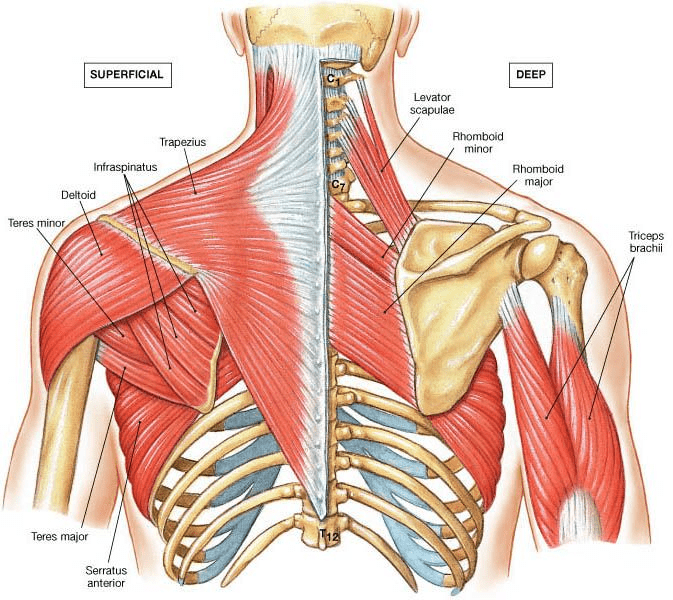
The back muscles are anatomically layered into superficial (extrinsic) and deep (intrinsic) muscles. Thoracic spine anatomy and upper back pain. It consists of three sections, the upper arm, forearm, and hand. It consists of seven vertebrae. Your upper arm bone (humerus), your shoulder blade (scapula), and your collarbone (clavicle). Feel the stretch in the back of your upper right arm and shoulder. The scapula is a large, flat, and somewhat triangular bone that sits between the humerus (upper arm bone) and collarbone. The upper back (and neck) plays an important role in shoulder function for one very simple reason. Shoulder mri assesses the following tendon and muscle structures: The shoulder is made up of two joints, the acromioclavicular joint and the glenohumeral joint. There are three sets of iliocostalis muscles: The upper trapezius, the part that goes across the tops of your shoulders, can elevate or bring up your shoulder girdle. The anatomy of the shoulder.
The cervical spine supports the weight and movement of your head and protects the nerves exiting your brain. The bones of the chest and upper back combine to form the strong, protective rib cage around the vital thoracic organs such as the heart and lungs. Anatomy upper back shoulder flashcards on quizlet. The shoulder joint, also known as the glenohumeral joint, is a ball and socket joint with the most extensive range of motion in the human body. Anatomy of the upper back muscles the neck consists of seven cervical vertebrae, the building blocks of the spine.
Anatomy Of Upper Back Muscles Anatomy Drawing Diagram from physiologicnyc.com These important muscles control many motions that involve moving the arms and head — such as throwing a ball, looking up at the sky, and raising your hand. The bone is surrounded and supported by a complex system of muscles that work together to help you move your arm. It connects with the collarbone. Surrounding the shoulder joint is the rotator cuff, which is a group of muscles and tendons (12). The shoulder joint, also known as the glenohumeral joint, is a ball and socket joint with the most extensive range of motion in the human body. The trapezius and latissimus dorsi muscles connect the upper limb to the vertebral column. Anatomy upper back shoulder flashcards on quizlet. Anatomy upper back shoulder with free interactive flashcards.
The muscle then courses up to your shoulder and attaches to your upper arm bone.
Connecting with the cervical spine above and the lumbar spine below, the thoracic spine runs from the base of the neck down to the abdomen. Injuries to these muscles can cause upper back pain. Thoracic spine anatomy and upper back pain. If you are curious about back anatomy and how to draw the back, follow to my tutorial on drawing the back at this link. Each block is separated by a disc that sits in between and each vertebra has a facet joint on either side. The bone is surrounded and supported by a complex system of muscles that work together to help you move your arm. It consists of seven vertebrae. It runs from the neck to the upper back. The trapezius and latissimus dorsi muscles connect the upper limb to the vertebral column. It's a vast area when you consider it encompasses the thoracic spine and. Place your right hand on your right shoulder. It helps stabilize the shoulder's movements. More commonly known as the shoulder blade, the scapula is a flat triangular bone located in the upper back.
The intrinsic back muscles are found deeper to the extrinsic muscles, separated from them by the thoracolumbar fascia. Powerful muscles that move the head and arms attach to these bones as well. Your lats are a major back muscle and mover of your shoulder joint. The extrinsic back muscles are located in the back, but act to produce movements of the shoulder and assist respiration. If you are curious about back anatomy and how to draw the back, follow to my tutorial on drawing the back at this link.
Body Anatomy Upper Extremity Bones The Hand Society from www.assh.org The shoulder is made up of 3 bones: Your upper arm bone (humerus), your shoulder blade (scapula), and your collarbone (clavicle). Shoulder mri assesses the following tendon and muscle structures: The shoulder joint is formed where the humerus (upper arm bone) fits into the scapula (shoulder blade), like a ball and. Connecting with the cervical spine above and the lumbar spine below, the thoracic spine runs from the base of the neck down to the abdomen. It runs from the neck to the upper back. It's a vast area when you consider it encompasses the thoracic spine and. Choose from 500 different sets of anatomy.
1) in the cervical area (iliocostalis cervicis), 2) in the upper back or thoracic area (iliocostalis thoracis), and 3) in the lumbar area (iliocostalis lumborum).
The extrinsic back muscles are located in the back, but act to produce movements of the shoulder and assist respiration. It consists of seven vertebrae. It connects with the collarbone. There are three sets of iliocostalis muscles: The cervical spine supports the weight and movement of your head and protects the nerves exiting your brain. Place your right hand on your right shoulder. Connecting with the cervical spine above and the lumbar spine below, the thoracic spine runs from the base of the neck down to the abdomen. The anatomy of the shoulder. The glenohumeral joint is where the ball (humeral head) and the socket (the glenoid) meet. It is the only spinal region attached to the rib cage. The thoracic spine is the longest region of the spine, and by some measures it is also the most complex. If you are curious about back anatomy and how to draw the back, follow to my tutorial on drawing the back at this link. It is responsible for stabilizing the upper arm bone, which sits in a shallow socket on the outer edge of the shoulder blade.
It consists of seven vertebrae upper back anatomy. Keeping your shoulders down and back, lift your right elbow up toward the ceiling to the point of tightness.


0 Komentar